Images in Infectious Diseases: Atypical manifestation of leprosy in a pediatric patient
Although leprosy is an endemic disease in Brazil, it remains underdiagnosed
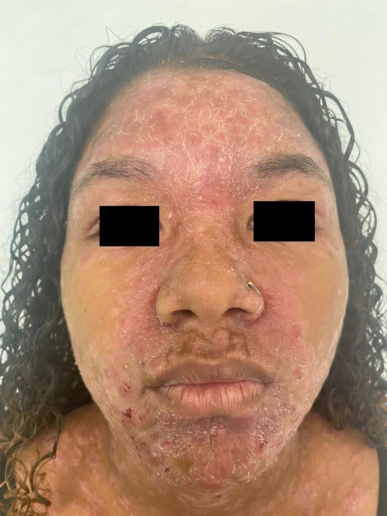
19/12/2024Erythematous-scaly plaques grouped in the frontal and central-facial region
Tavares GC – Atypical pediatric’s leprosy
Gabriel Castro Tavares[1]
[1]. Instituto de Puericultura e Pediatria Martagão Gesteira, Serviço de Dermatologia, Rio de Janeiro, RJ, Brasil.
Corresponding author: Gabriel Castro Tavares. e-mail: gabrielcastrotavares@gmail.com
Authors’ contribution
GCT: Conception and design of the study, Acquisition of data, Drafting the article, Final approval of the version to be submitted.
Conflict of Interest
There was no conflict of interest.
- Financial Support
There was no financial support.
- Orcid
Gabriel Castro Tavares: https://orcid.org/0000-0001-9914-2428
Received 23 August 2024 – Accepted 20 September 2024
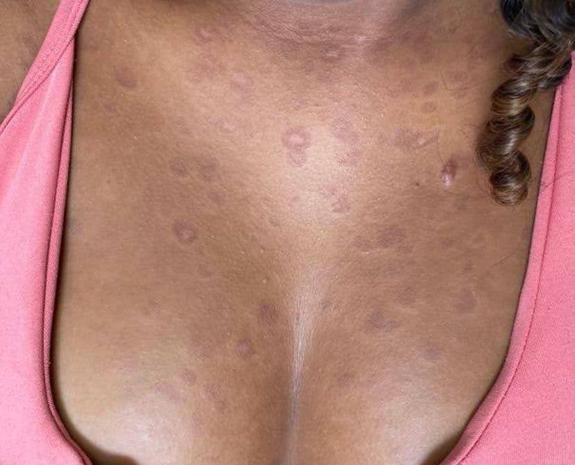
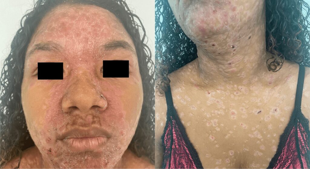
A teenager, 14 years old, reported the progressive appearance of skin lesions for two months, which was unassociated with other symptoms. Physical examination revealed erythematous and scaly plaques, some with a crusted center and oval shape, with a diffuse and symmetrical distribution located on the trunk, upper limbs, and face, grouped in the frontal and center-facial regions (Figures 1 and 2). There was no change in the peripheral nerves. An incisional biopsy was performed on a trunk lesion compatible with reactional leprosy. A diagnosis of dimorphic leprosy associated with a reverse reaction was reached after clinical-pathological correlation. The treatment consisted of oral prednisone (1 mg/kg/day) combined with rifampicin, clofazimine, and dapsone for 12 months until clinical improvement of the reaction was observed. The clinical appearance of the skin lesions changed after one week of prednisone treatment. Physical examination revealed erythematous brownish-to-violet plaques, non-scaling, with infiltrated edges and an annular shape (Figure 3), compatible with the classic picture of leprosy after improvement in the reaction. Although leprosy is an endemic disease in Brazil, it remains underdiagnosed. It is caused by Mycobacterium leprae and primarily affects the skin and peripheral nerves, causing deformities and functional disabilities if not treated early. Although the diagnosis is based on clinical findings, the inflammatory nature of the leprosy reaction modifies the classic presentation of the disease1-3, so a histopathological examination is essential to confirm the diagnosis and initiate treatment early.
References
- Peng J, Sun P, Wang L, Wang H, Long S, Yu MW. Leprosy among new child cases in China: Epidemiological and clinical analysis from 2011 to 2020. PLoS Negl Trop Dis. 2023;17(2):e0011092.
- Putri AI, de Sabbata K, Agusni RI, Alinda MD, Darlong J, de Barros B, Walker SL, Zweekhorst MBM, Peters RMH. Understanding leprosy reactions and the impact on the lives of people affected: An exploration in two leprosy endemic countries. PLoS Negl Trop Dis. 2022;16(6):e0010476.
- Reza NR, Kusumaputro BH, Alinda MD, Listiawan MY, Thio HB, Prakoeswa CRS. Pediatric Leprosy Profile in the Postelimination Era: A Study from Surabaya, Indonesia. Am J Trop Med Hyg. 2022;106(3):775-8.

FIGURE 1: Erythematous and scaly plaques with a crusted center and oval shape located on the anterior chest.

FIGURE 2: Erythematous-scaly plaques grouped in the frontal and central-facial region.

FIGURE 3: Erythematous brownish to violet plaques, non-scaling, with infiltrated edges and annular shape located on the anterior chest.


