Multiple lung nodules with the halo sign due to syphilis
Patient met the diagnostic criteria of pulmonary syphilis, which include typical findings of syphilis in other organs, positive serological findings, imaging abnormalities that cannot be explained by other conditions, and response to syphilis treatment
08/08/2023
Pulmonary syphilis in a 26-year-old man. (A–C) An axial chest CT scan showing multiple nodules in both lungs, some with peripheral ground-glass halos, constituting the halo sign (arrows). (D–F) A chest CT scan performed 2 weeks after receiving penicillin G showing the resolution of all lesions
Corrêa DG et al. – Pulmonary syphilis
Diogo Goulart Corrêa[1],[2], Rodrigo Paulino[1] and Roberto Mogami[1]
[1]. Universidade do Estado do Rio de Janeiro, Departamento de Medicina Interna, Disciplina de Radiologia, Rio de Janeiro, RJ, Brasil.
[2]. Clínica de Diagnóstico por Imagem/DASA, Departamento de Radiologia, Rio de Janeiro, RJ, Brasil.
Corresponding author: Diogo Goulart Corrêa. e-mail: diogogoulartcorrea@yahoo.com.br
Authors’ contribution
DGC: study conception, initial drafting of the manuscript, and review of the literature.
RP: data acquisition, analysis and interpretation of data, and critical revision of the manuscript for intellectual content.
RM: data acquisition, analysis and interpretation of data, and critical revision of the manuscript for intellectual content.
All authors approved the final version of the manuscript and agree to be accountable for all aspects of the work.
Conflict of Interest
The authors declare that they have no conflict of interest.
Financial Support
None.
Orcid
Diogo Goulart Corrêa: https://orcid.org/0000-0003-4902-0021
Rodrigo Paulino: https://orcid.org/0009-0001-4002-0001
Roberto Mogami: https://orcid.org/0000-0002-7610-2404
A 26-year-old man presented with fever, a maculopapular rash on the chest and face, oral and genital ulcers, and cough for 2 weeks. Serological testing revealed positivity for human immunodeficiency virus, with a viral load of 18,218 copies/mL and a CD4 T lymphocyte count of 880 cells/mm3. The Venereal Disease Research Laboratory test revealed serum positivity (1:512), and the fluorescent treponemal antibody absorption test was reactive. Chest computed tomography (CT) revealed multiple nodules in both lungs, some with peripheral ground-glass halos. The patient was treated with penicillin G. A chest CT performed 2 weeks after treatment showed resolution of all lesions (Figure 1).
Pulmonary involvement may occur in the secondary stage of syphilis due to the hematogenous spread of Treponema pallidum or in the tertiary stage with gummas and/or fibrotic lesions1. Our patient met the diagnostic criteria of pulmonary syphilis, which include typical findings of syphilis in other organs, positive serological findings, imaging abnormalities that cannot be explained by other conditions, and response to syphilis treatment2. However, pulmonary syphilis can occur without the typical cutaneous lesions3. The imaging findings of pulmonary syphilis include multiple nodules2 and/or masses3 simulating primary lung neoplasia or metastases, and abscesses1 with or without mediastinal lymph node enlargement. 18F fluorodeoxyglucose (18F-FDG) positron emission tomography–CT may show intense 18F-FDG uptake by the lesions2. Although rarely reported, pulmonary syphilis may be under-recognized since chest imaging is not routinely performed. Therefore, syphilis should be included in the differential diagnosis of patients with respiratory symptoms and unexplained lung nodules or masses3.
Acknowledgments
None.
References
1 – Visuttichaikit S, Suwantarat N, Apisarnthanarak A, Damronglerd P. A case of secondary syphilis with pulmonary involvement and review of the literature. Int J STD AIDS. 2018;29(10):1027-32. Available from: https://doi.org/10.1177/0956462418765834
2 – Kim SJ, Lee JH, Lee ES, Kim IH, Park HJ, Shin C, et al. A case of secondary syphilis presenting as multiple pulmonary nodules. Korean J Intern Med. 2013;28(2):231-5. Available from: https://doi.org/10.3904/kjim.2013.28.2.231
3 – Florencio KBV, Costa ADD, Viana TCM, Gomes DCA, Gouveia PADC. Secondary syphilis with pulmonary involvement mimicking lymphoma: a case report. Rev Soc Bras Med Trop. 2019;52:e20190044. Available from: https://doi.org/10.1590/0037-8682-0044-2019
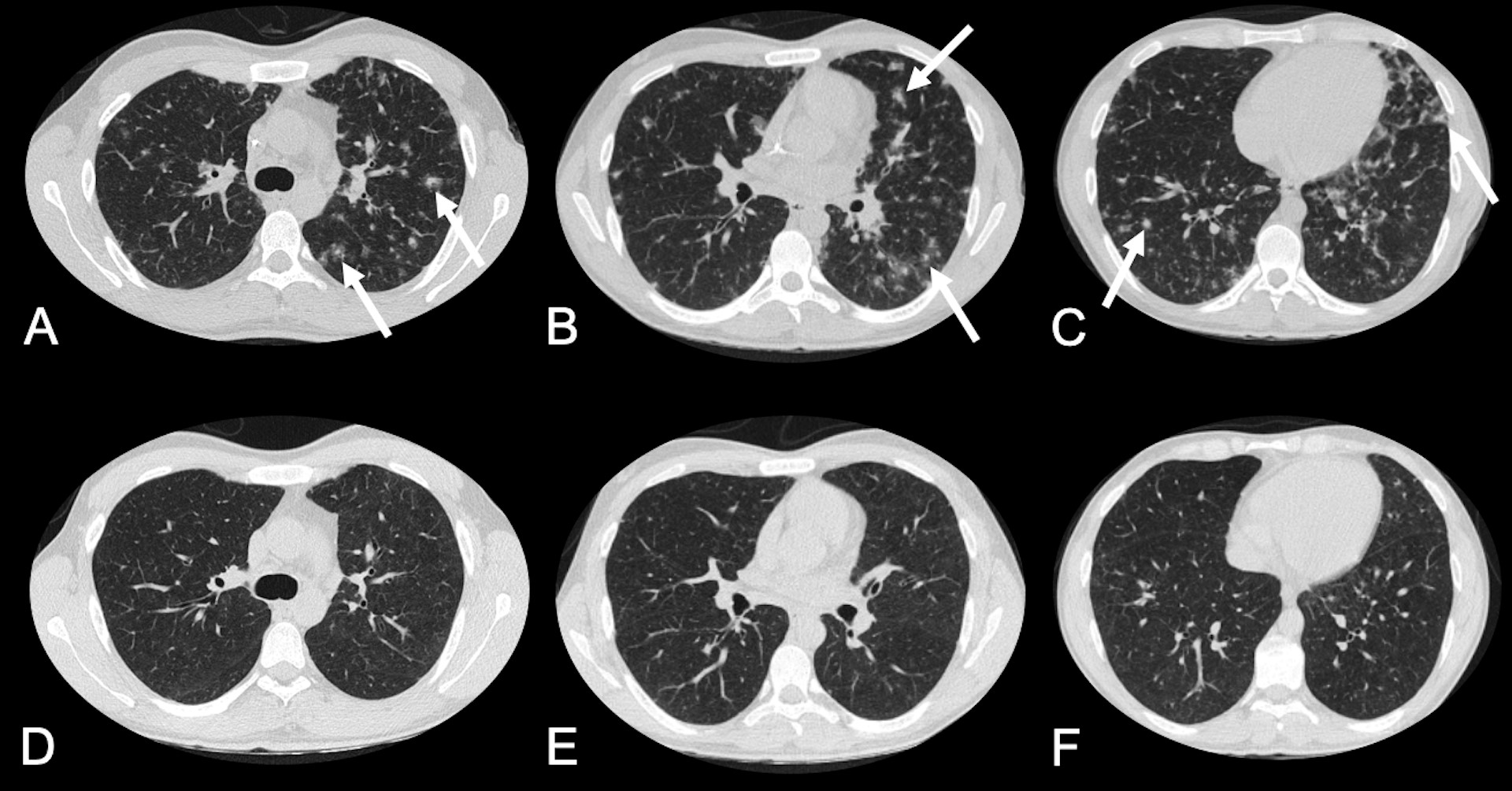
FIGURA 1: Pulmonary syphilis in a 26-year-old man. (A–C) An axial chest CT scan showing multiple nodules in both lungs, some with peripheral ground-glass halos, constituting the halo sign (arrows). (D–F) A chest CT scan performed 2 weeks after receiving penicillin G showing the resolution of all lesions
**Esta reportagem reflete exclusivamente a opinião do entrevistado.**