
Gökhan Polat[1], Büşra Şahin Toprak[1] and Enes Yılmaz[1]
[1]. Department of Radiology, Medical Faculty, Kutahya Health Sciences University, Kutahya, Turkey.
Correspondence: Dr. Büşra Şahin Toprak. e-mail: [email protected]
Financial Support: The authors received no financial support for the research and/or authorship of this article.
Conflict of Interest: The authors declare that they have no conflict of interest to the publication of this article.
ORCID iDs
Gökhan Polat 0000-0002-9184-8730
Büşra Şahin Toprak 0000-0002-2145-5544
Enes Yılmaz 0000-0002-0279-994X
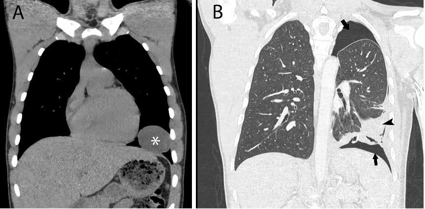
A 17-year-old male patient presented to our hospital four months ago with complaints of cough, difficulty in breathing, and a stabbing sensation. A non-contrast thoracic computed tomography (CT) scan revealed a well-defined cystic lesion in the lower lobe of the left lung (Figure 1A). Oral albendazole was initiated for hydatid cyst (HC) treatment. In the fourth month of treatment, the patient presented with hemoptysis and dyspnea. A posteroanterior chest radiograph revealed extensive pneumothorax necessitating chest tube placement. A thoracic CT scan confirmed rupture of the cyst in the left lung (Figure 1B).
The hydatid disease belongs to the Echinococcus species that primarily affects the liver and lungs. HC progresses slowly and can remain asymptomatic. Pulmonary HC typically become inactive due to fibrosis 5–9 months after treatment1. Albendazole has been used to treat HC for approximately five decades. Additionally, they facilitate cyst treatment by activating certain cellular death mechanisms in the germinative membrane.² Some studies have reported ruptures as a complication of albendazole therapy. Notably, the study by Kürkçüoğlu et al.² provides examples of such cases. Although these compounds are effective therapeutic agents for hydatid cysts, they are associated with a non-negligible risk of complications.
or contributions
GP: Writing—review & editing; BŞT: Writing—review & editing supporting; EY: Writing—review & editing.
References
- Daimari R, Oyekunle AA, Ocampo C. Ruptured Pulmonary Hydatid Cyst Following the Use of Albendazole. Am J Trop Med Hyg. 2018;98(6):1555-6.
- Kurkcuoglu IC, Eroglu A, Karaoglanoglu N, Polat P. Complications of albendazole treatment in hydatid disease of lung. Eur J Cardiothorac Surg. 2002;22(4):649-50.
FIGURE 1: Non-contrast coronal thoracic computed tomography (A) showing a well-defined cystic lesion in the lower lobe of the left lung in the mediastinal window (asterisk). The cyst density measured to be 10 HU. Contrast-enhanced coronal thoracic computed tomography (B) demonstrating air densities consistent with pneumothorax between the pleural layers in the parenchymal window (black arrows). Additionally, marked wall irregularity and pleuropulmonary fibroatelectatic changes with volume loss are observed at the previously identified location of the cyst in the lower lobe of the left lung (black arrowhead).