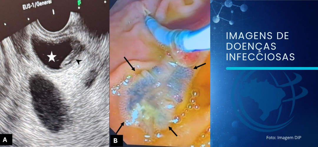
EUS (A) shows echogenic, flat-shaped material (arrowhead) in the dilated CBD (white star), and ERCP (B) demonstrates a F. hepatica worm (black arrows)
Gündoğdu E- Pancreatitis caused by Fasciola hepatica
Elif Gündoğdu[1], Berrin Yalınbaş Kaya[2] and Melikşah Yüksel[3]
[1]. Eskişehir Osmangazi University, Faculty of Medicine, Department of Radiology, Eskişehir, Turkey.
[2]. University of Health Sciences, Ministry of Health Eskisehir City Hospital, Department of Gastroenterology, Eskişehir, Turkey.
[3]. Eskişehir Osmangazi University, Faculty of Medicine, Department of Gastroenterology, Eskişehir, Turkey.
Corresponding author: Dr. Elif Gündoğdu. E-mail: [email protected]
Conflict of Interest Statement: The authors declare no conflicts of interest.
Financial Support: None.
Editor-in-Chief: Prof. Dalmo Correia Filho
Orcid
Elif Gündoğdu: https://orcid.org/0000-0002-1729-6958
Berrin Yalınbaş Kaya: https://orcid.org/0009-0001-8366-9601
Melikşah Yüksel : https://orcid.org/0009-0004-3667-5832
A 62-year-old male presented with abdominal pain radiating to the back along with nausea and vomiting. Examination revealed abdominal distension and epigastric tenderness, and the patient’s vital signs were normal. Laboratory tests showed elevated levels of alanine aminotransferase, aspartate aminotransferase, gamma-glutamyltransferase, amylase, and lipase, along with eosinophilia (93 U/L, 102 U/L, 78 U/L, 1,292 U/L, 600 U/L, and 0.40×10³/μL, respectively). The patient had experienced an episode of acute pancreatitis one year earlier, with recurrent admissions for similar symptoms. Magnetic resonance cholangiopancreatography demonstrated findings of acute edematous pancreatitis, with flat-shaped, facetless signal loss in the gallbladder and dilatation of the common bile duct (CBD) with similar flat signal loss (Figure 1). A retrospective review of computed tomography scan performed one year earlier revealed millimeter-sized, clustered, tubular multicysts in the subcapsular areas of the liver parenchyma (Figure 2). With a preliminary diagnosis of infection with Fasciola hepatica, endoscopic ultrasonography and endoscopic retrograde cholangiopancreatography (ERCP) were performed, and an F. hepatica was extracted from the CBD (Figure 3). F. hepatica is a flatworm that infects the liver and biliary tract of humans1. Transmission occurs through the ingestion of water or food contaminated with metacercariae. After excysting in the intestine, the parasite penetrates the peritoneal cavity and liver capsule, migrates through the parenchyma and finally in the biliary tract2. The clinical presentation is variable and can manifested as abdominal pain, fever, and jaundice3. Acute pancreatitis is rarely observed3. In patients presenting with recurrent acute pancreatitis, facetless signal loss and eosinophilia, infection with F. hepatica should be suspected. ERCP is essential for confirmation and treatment.
ACKNOWLEDGMENTS
The authors of the manuscript have no acknowledgments.
AUTHORS’ CONTRIBUTION
EG: Conception and design of the study, Acquisition of data, Analysis and interpretation of data, Drafting the article, Final approval of the version to be submitted. BYK: Acquisition of data, Analysis and interpretation of data, Final approval of the version to be submitted. MY: Acquisition of data, Analysis and interpretation of data, Final approval of the version to be submitted.
REFERENCES
- Altintas E, Sezgin O, Tombak A, Ucbilek E. Fasciola hepatica-induced acute pancreatitis: report of two cases and review of the literature. Turk J Gastroenterol. 2010;21(2):183–7.
- Huy DQ, Khai NX, Minh NT, Tien TD, Yen TH, Duc NM. Acute edematous pancreatitis caused by Fasciola hepatica infection: A rare case report. Radiol Case Rep. 2023;18(9):2907-10.
- Bandaru SS, Rawla P, Vellipuram AR. Review of infectious etiology of acute pancreatitis. Gastroenterol Res. 2017;10(3):153–8.
FIGURE 1: Coronal MR image demonstrates edematous pancreatitis and peripancreatic fluid (arrowheads), with flat, facetless signal loss in the gallbladder (black arrow), and dilatation of the CBD with flat signal loss (blue arrow).
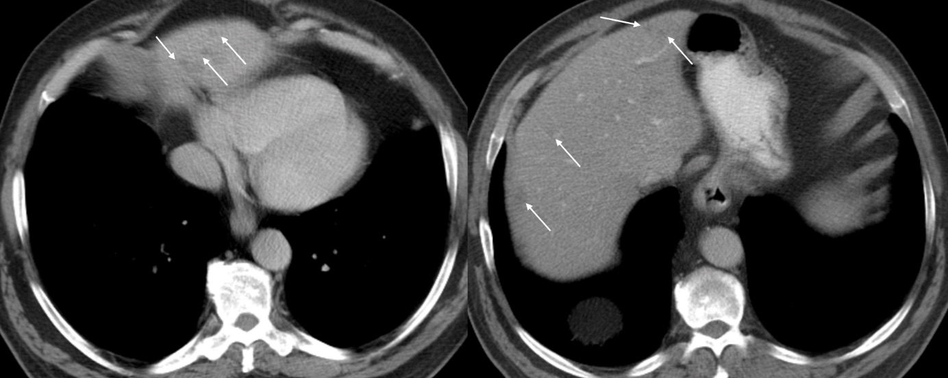
FIGURE 2: CT demonstrates millimetric to subcentimetric multicysts in the subcapsular areas of the liver parenchyma (white arrows).
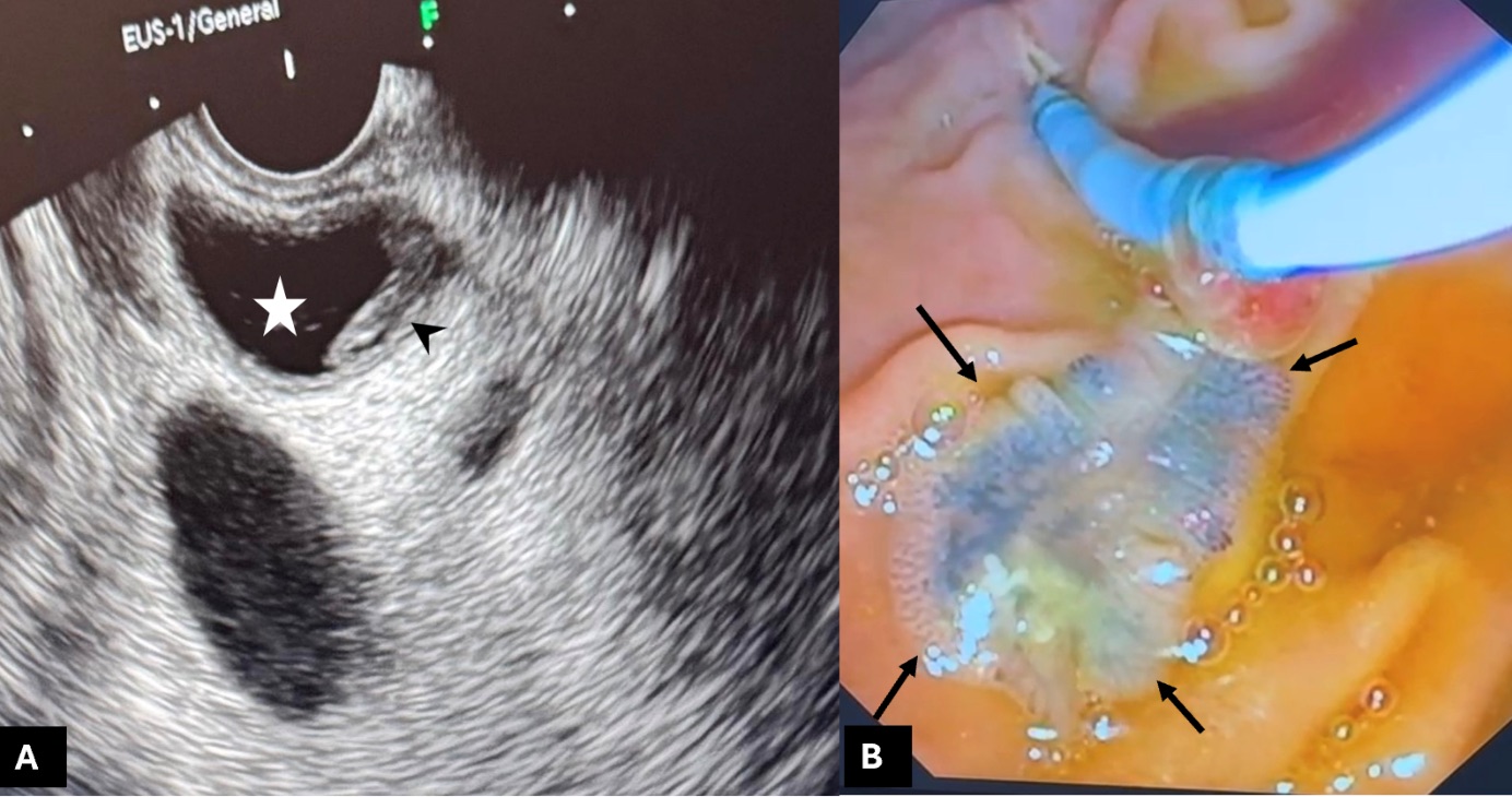
FIGURE 3: EUS (A) shows echogenic, flat-shaped material (arrowhead) in the dilated CBD (white star), and ERCP (B) demonstrates a F. hepatica worm (black arrows).