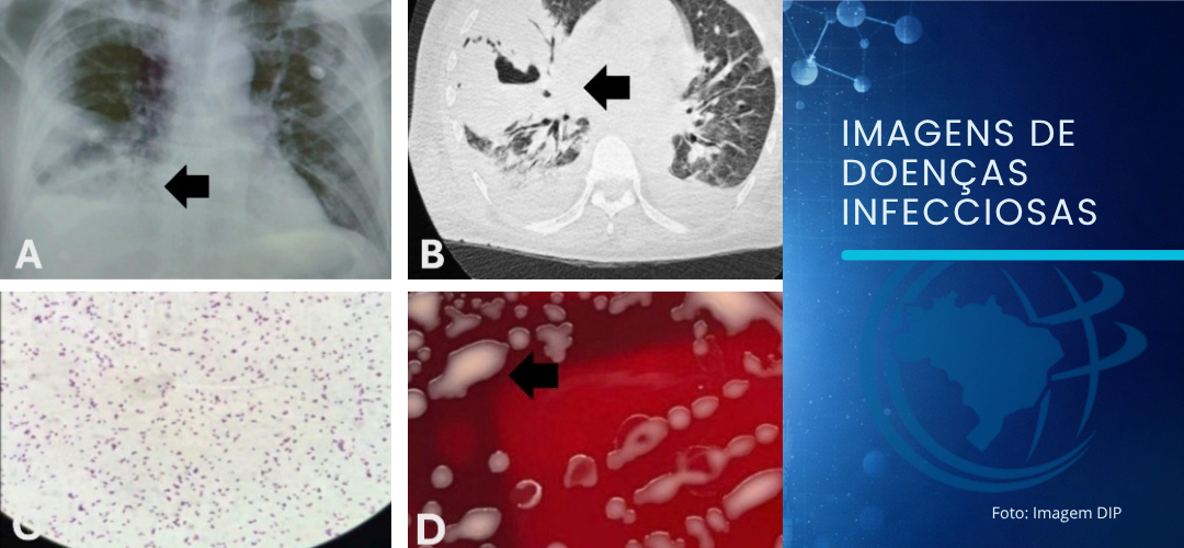
Imaging showed consolidation and cavitation with air-fluid levels in the middle lobe. Gram-positive coccobacilli were identified, and cultures confirmed Rhodococcus equi
Silva EFB– Pulmonary infection caused by Rhodococcus equi
Eduardo Felipe Barbosa Silva[1], Alessandra Fracaro Camargo[1] and Lilian Alves Rocha[2]
[1].Hospital Regional da Asa Norte, Unidade de Doenças Torácicas, Broncoscopia, Brasilia, DF, Brasil.
[1]. Laboratório Central de Saúde Pública do Distrito Federal, Brasilia, DF, Brasil.
Corresponding author: Eduardo Felipe Barbosa Silva. E-mail: [email protected]
Conflict of Interest Statement: The authors declare no conflicts of interest.
Financial Support: None.
Editor-in-Chief: Manoel Otavio da Costa Rocha
ORCID
Eduardo Felipe Barbosa Silva: https://orcid.org/0000-0001-9610-6827
Alessandra Fracaro Camargo: https://orcid.org/0009-0008-6203-5694
Lilian Alves Rocha: https://orcid.org/0009-0009-1591-5784
A 47-year-old man presented with fever, chest pain, cough, dyspnea, and weight loss (11 kg) for three months. Chest radiography and computed tomography (Figure 1A, B) revealed consolidation with air bronchograms and central cavitation with fluid accumulation, forming air-fluid levels in the middle lobe. Reactive human immunodeficiency virus (HIV) serology was performed, and the CD4 count was 22 cells/mm3. Blood and bronchoalveolar lavage cultures demonstrated growth of Rhodococcus equi on blood agar (Figure 1C, D). Despite antibiotic therapy, the patient developed septic shock and died.
R. equi, the causative agent of rhodococcosis, is a gram-positive, aerobic, and partially acid-fast coccobacillus1-3. It is an opportunistic pathogen that affects patients with altered cellular immunity, including solid-organ and hematological transplant recipients, and patients with HIV1. Approximately 80% of cases of rhodococcosis occur in patients with HIV3. Its natural reservoir is soil, and inhalation is the most common route of infection1,3. The lung is the most commonly affected organ in individuals with HIV, whereas extrapulmonary forms are mainly in patients without HIV1-3.
Clinically, R. equi presents as subacute or chronic cavitary pneumonia3. The main pulmonary imaging finding is consolidation with cavities, with or without fluid levels2,3. Microbiological diagnosis is based on direct examination (Gram, Ziehl–Neelsen, and Kinyoun staining) and culture on blood agar and Lowenstein–Jensen media1,3. Treatment includes combination antibiotic therapy (macrolides, rifampicin, fluoroquinones, aminoglycosides, glycopeptides, and carbapenems)3.
In patients with HIV and a CD4 count <200 cells/mm3 and subacute/chronic cavitary lung injury, rhodococcosis should be considered in the differential diagnosis.
Acknowledgments
None.
Authors’ contribution
EFBS: Performed the bronchoscopy examination and wrote the medical article; AFC: Performed the bronchoscopy examination and wrote the medical article; LAR: Microbiological diagnosis.
REFERENCES
- Severo LC, Ritter P, Petrillo VF, Dias CAG, Porto NS. Pulmonary Rhodococcus equi infection: report of the first two Brazilian cases. J Pneumol. 2001;27(3):158-62.
- Marchiori E, de Mendonça RG, Capone D, de Cerqueira EM, Souza Júnior AS, Zanetti G, et al. Rhodococcus equi infection in acquired immunodeficiency syndrome. Computed tomography aspects. J Bras Pneumol. 2006;32(5):405-9.
- Vechi HT, Oliveira ETG, Freitas MR, Rossi F, Britto MHMF, Alves MDM. Chronic cavitary pneumonia by Rhodococcus equi in a highly prevalent tuberculosis country: a diagnosis challenge. Rev Inst Med Trop Sao Paulo. 2018;60:e74.
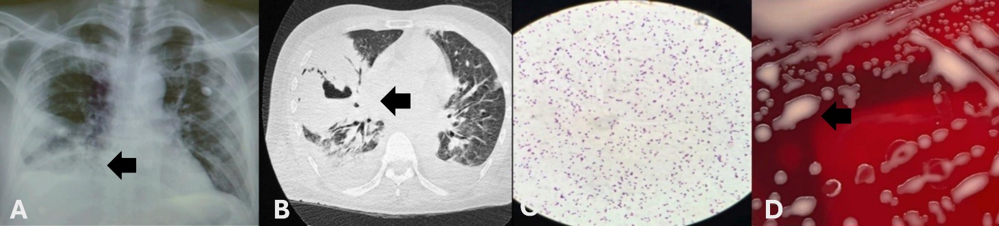
FIGURE 1: (A and B) Chest radiography and computed tomography showing consolidation and central cavitation with fluid accumulation, forming air-fluid levels in the middle lobe. (C) Gram-positive coccobacillus on direct examination. (D) culture media showing growth of Rhodococcus equi on blood agar.