
Pulmonary mucormycosis is a rare but highly lethal opportunistic infection in solid-organ transplant recipients
Tamiozzo MM et al. – Pulmonary mucormycosis cavity in kidney transplant
Marina Manica Tamiozzo[1], Leticia Dalmolin[1] and Mariana Manica Tamiozzo[2]
[1]. Universidade Federal de Santa Maria, Centro de Ciências da Saúde, Santa Maria, RS, Brasil.
[2]. Hospital Universitário de Santa Maria, Departamento de Radiologia e Diagnóstico por Imagem, Santa Maria, RS, Brasil.
Corresponding author: Miss. Marina Manica Tamiozzo. Address: 1000 Roraima Avenue, University Campus, Camobi District, Santa Maria, RS, 97105-900, Brazil. Phone number: 55 99103-7588. E-mail: [email protected]
Conflict of Interest Statement: The authors declare no conflicts of interest.
Financial Support: None.
Editor-in-Chief: Prof. Dalmo Correia Filho
ORCID
Marina Manica Tamiozzo: https://orcid.org/0009-0008-0765-2948
Leticia Dalmolin: https://orcid.org/0009-0008-2515-8652
Mariana Manica Tamiozzo: https://orcid.org/0000-0003-1984-3721
A 67-year-old man with chronic kidney disease receiving immunosuppressive therapy after kidney transplantation was hospitalized for diabetic ketoacidosis. Chest radiography at admission demonstrated a prominent pulmonary cavitation in the left lung (Figure 1). Subsequently, computed tomography of the chest revealed a large cavitary lesion with thick walls and multiple internal septations in the left upper lobe, measuring approximately 6.0 × 5.0 cm. Faint adjacent ground-glass opacities and small bilateral pulmonary nodules were also observed.
Given the patient’s severe immunosuppression, an infectious process, including a lung abscess, tuberculosis, and fungal infection, was suspected as the differential diagnosis. Culture results and histopathological analysis of the lesion ultimately revealed aseptate hyaline hyphae with right-angle branching, consistent with mucormycosis. However, despite diagnostic efforts, the patient died during hospitalization from complications of end-stage renal disease.
Pulmonary mucormycosis is a rare but highly lethal opportunistic infection in solid-organ transplant recipients1-2. This case emphasizes the critical importance of recognizing radiological features suggestive of pulmonary mucormycosis, such as thick-walled cavities with internal septations, to enable prompt targeted antifungal therapy and timely surgical intervention3-4
Authors’ contributions
MMT: Conceptualization, writing, reviewing, drafting, editing, and supervision of the manuscript; LD: Conceptualization, writing, reviewing, drafting, editing, and supervision of the manuscript; MAT: Reporting of the computed tomography findings, conceptualization, writing, reviewing, drafting, editing, and supervision of the manuscript.
Acknowledgments
We offer our deepest thanks to the institutions that provided technical support for the development and implementation of this study.
References
- Navarro Vergara DI, Barragán Pola G, Bonifaz A, Núñez Pérez-Redondo C, Choreño García O, Cicero Sabido R, et al. Pulmonary mucormycosis in a patient with kidney transplant and uncontrolled haemoptysis. Rev Iberoam Micol. 2017;34(4):233-6.
- Boán J, Fernández-Ruiz M, Aparicio-Minguijón E, Aguado JM. The unnoticed threat: clinical characteristics, risk factors, and outcome of mucormycosis in solid-organ transplantation. J Fungi. 2025;11:853.
- Hase A, Dalal A, Bhanushali A, et al. Successful management of pulmonary mucormycosis presenting as round pneumonia by lung resection in a kidney-transplant recipient. Saudi J Kidney Dis Transpl. 2023;34(Suppl 1):S226-9.
- Baddley JW, Forrest GN; American Society of Transplantation Infectious Diseases Community of Practice. Cryptococcosis, blastomycosis, and mucormycosis in solid organ transplant recipients. Am J Transplant. 2019;19(Suppl 3):254-65.
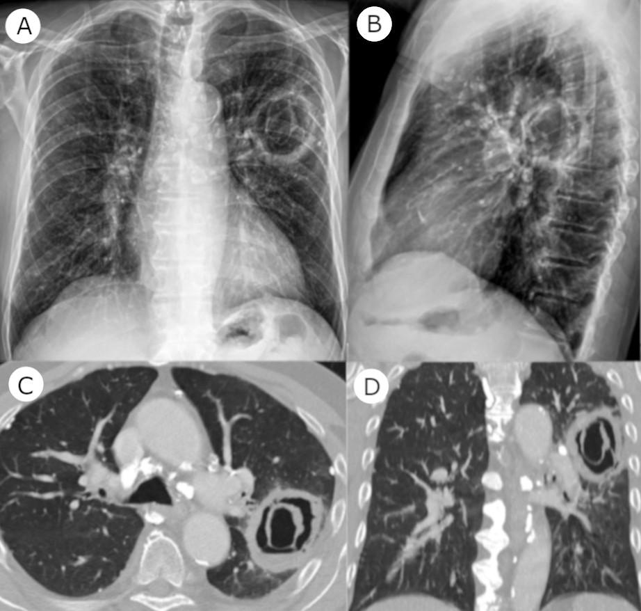
FIGURE 1: (A) Anteroposterior chest radiograph showing a cavitary lesion in the left upper lobe. (B) Lateral chest radiograph confirming the lesion’s presence and location. (C) Axial chest computed tomography showing a thick-walled cavitary lesion with internal septations in the left upper lobe, corresponding to the coronal view shown in (D).