Santos LB et al. Cutaneous infection by Mycobacterium chelonae
Lucas Benício dos Santos[1], Rafael Henrique Bento Elizeu[2], Lida Jouca de Assis Figueredo[2], Élida Aparecida Leal[3] and Silvana Spíndola de Miranda[2]
[1]. Universidade Federal de Minas Gerais, Faculdade de Medicina, Departamento de Propedêutica Complementar. Belo Horizonte, MG, Brasil.
[2]. Universidade Federal de Minas Gerais, Faculdade de Medicina, Departamento de Clínica Médica, Belo Horizonte, MG, Brasil.
[3]. Fundação Ezequiel Dias, Serviço de Doenças Bacterianas e Fúngicas. Belo Horizonte, MG, Brasil.
Correspondence: Lucas Benício dos Santos. E-mail: lucasbenicio2010@gmail.com
Ethics: Report from the Ethics Committee of the Federal University of Minas Gerais, approved under protocol number 6.682.998 (CAAE 76360223.6.0000.5149).
Financial Support: None
Conflict of Interest: The authors declare no conflicts of interest.
Editor: Manoel Otavio da Costa Rocha
Lucas Benício dos Santos. ORCID: https://orcid.org/0000-0003-2242-8414
Rafael Henrique Bento Elizeu. ORCID: https://orcid.org/0009-0008-9019-1555
Lida Jouca de Assis Figueredo. ORCID: https://orcid.org/0000-0001-5355-0784
Élida Aparecida Leal. ORCID: https://orcid.org/0000-0002-5998-7335
Silvana Spíndola de Miranda. ORCID: https://orcid.org/0000-0001-7245-4472
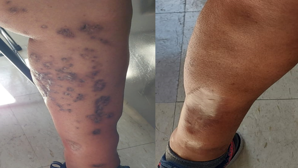
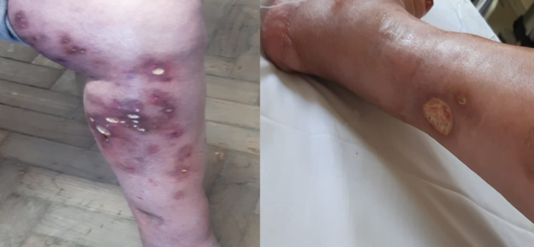
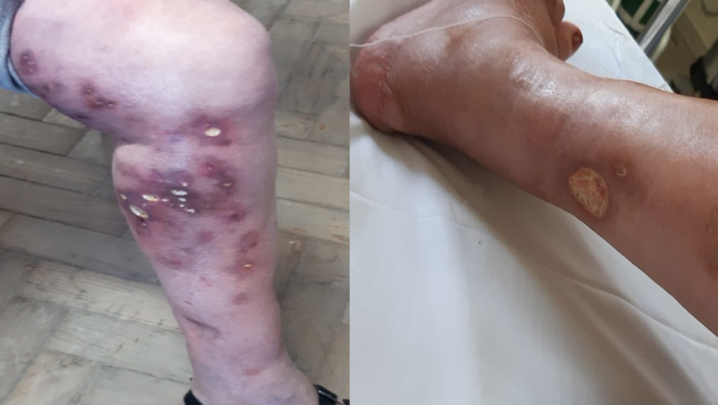
A 55-year-old woman with type II diabetes, a former smoker, and a history of immunosuppression was treated with methotrexate for pyoderma gangrenosum between 2009 and 2013. During this period, the lesions exhibited intermittent improvement followed by worsening. In 2021, she was referred to the Tuberculosis Reference Clinic for a skin biopsy with positive bacilloscopy and a positive culture for rapidly growing mycobacteria. At that time, hyperchromic, hyperemic, and edematous macules and circular ulcers with purulent exudates that drained spontaneously were observed on her lower limbs (Figure 1). The identification tests confirmed the presence of Mycobacterium chelonae. The treatment lasted 18 months and initially included amikacin, clarithromycin, and moxifloxacin. Sensitivity testing showed resistance to moxifloxacin, which was replaced with clofazimine. Bacilloscopy and mycobacterial cultures were performed and the wounds tested negative for draining secretions. By the end of the treatment, the lesions were reduced, which were accompanied by hyperchromic macules, some of which had dry crusts (Figure 2).
Nontuberculous mycobacteria are ubiquitous and can cause diverse conditions, from asymptomatic colonization to infections1,3. Diseases caused by M. chelonae are often associated with invasive procedures4. In this case, the microorganism was considered opportunistic and favored by prior immunosuppression3. Although it is an uncommon pathogen5, considering its possibility in the absence of a response to initial therapy is crucial. Careful attention to the differential diagnosis of atypical lesions and performing mycobacteriological tests for accurate and timely diagnosis and treatment are necessary1,2.
ACKNOWLEDGMENTS
We thank Cláudio José Augusto, the Bacterial and Fungal Doenças Service of the Ezequiel Dias Foundation (FUNED) at the Microbacterium Research Laboratory of the Faculty of Medicine of UFMG, and the Medical and Statistical Archive Service of the Hospital das Clínicas of UFMG for their support in conducting this project.
AUTHORS’ CONTRIBUTIONS
LBS: conceptualization; data curator; formal analysis; investigation; methodology; project administration; validation; visualization; redaction of the original manuscript; drafting-review and editing. RHBE: conceptualization; dice curator; investigation; methodology; visualization; redaction of the original manuscript. LJAF: conceptualization; formal analysis; methodology; validation; drafting-review and editing. EAL: formal analysis; validation; drafting-review and editing. SSM: conceptualization; formal analysis; methodology; resources; supervision; validation; drafting-review and editing.
References:
- Ministério da Saúde (MS). Secretaria de Vigilância em Saúde. Departamento de Doenças de Condições Crônicas e Infecção de Transmissão Sexual. Recomendações para o diagnóstico e tratamento das doenças causadas por micobactérias não tuberculosas no Brasil. 1ª edição. Brasília – DF: MS; 2021 [cited Oct 01 2025]. Available from: https://www.gov.br/aids/pt-br/central-de-conteudo/publicacoes/2021/recomendacoes-para-o-diagnostico-e-tratamento-das-doencas-causadas-por-micobacterias-nao-tuberculosas-no-brasil/view
- Fundação Ezequiel Dias (FUNED). Instituto Octávio Magalhães. Serviço de Doenças Bacterianas e Fúngicas – Laboratório de Micobacterioses. Recomendações para o diagnóstico e teste de sensibilidade para micobactérias não tuberculosas (MNT) no estado de Minas Gerais, 2020 [cited Oct 01 2025]. Avaliable from: http://www.funed.mg.gov.br/wp-content/uploads/2020/10/RECOMENDACOES-DIAGNOSTICO-MICOBACTERIAS-MNT.pdf
- Haworth CS, Banks J, Capstick T, et al. British Thoracic Society guidelines for the management of non-tuberculous mycobacterial pulmonary disease (NTM-PD). Thorax. 2017;72(Suppl 2):ii1-ii64 [cited Oct 01 2025]. Avaliable from: https://thorax.bmj.com/content/72/Suppl_2/ii1.long
- Höfling-Lima AL, Freitas D, Sampaio JLM, Leão SC, Contarini P. In Vitro Activity of Fluoroquinolones Against Mycobacterium abscessus and Mycobacterium chelonae Causing Infectious Keratitis After LASIK in Brazil. Cornea. 2025;24(6):p 730-734, [cited Oct 01 2025]. Avaliable from: https://pubmed.ncbi.nlm.nih.gov/16015094/
- Barboza GCS, Almeida IN, Santos LBD, Augusto CJ, Leal ÉA, Pádua CAM, et al. Nontuberculous mycobacteria in patients of a specialty hospital. Rev Inst Med Trop Sao Paulo. 2023;30;65:e42. Avaliable from: https://doi.org/10.1590/S1678-9946202365042