Imagem Case Report
Dendrogram with strains of Vibrio cholerae O1 and non-O1, where two groups stand out (V. cholerae O1 and V. cholerae non-O1)
Barberino MGMA et al. – Suspected cholera in Brazil after 18 years.
Maria Goreth Matos de Andrade Barberino[1],[2], Ana Verena Almeida Mendes [1],[2],[3], Gabriela Noronha Marques[1], Camila Araújo de Lorenzo Barcia[1], Marcela Almeida Muhana[4], Aline Macedo Carvalho Freitas[4], Mariana Leal de Souza Mercês[4], Ênio Silva Soares[4], Lázaro José Rodrigues de Souza[4], Arabela Leal e Silva de Mello[5], Felicidade Mota Pereira[5], André Felipe das Mercês Santos[6], Dália dos Prazeres Rodrigues[6], Guilherme de Sousa Ribeiro[7],[8] and Cristiane Wanderley Cardoso[4],[8].
[1]. Hospital São Rafael, Salvador, BA, Brasil.
[2]. Instituto D’OR de Pesquisa e Ensino (IDOR), Salvador, BA, Brasil.
[3]. Escola Bahiana de Medicina e Saúde Pública, Salvador, BA, Brasil.
[4]. Secretaria Municipal de Saúde de Salvador, Centro de Informações Estratégicas em Vigilância em Saúde, Salvador, BA, Brasil.
[5]. Secretaria de Saúde do Estado da Bahia, Laboratório Central do Estado da Bahia, Salvador, BA, Brasil.
[6]. Fundação Oswaldo Cruz, Instituto Oswaldo Cruz, Laboratório de Enterobactérias, RJ, Brasil.
[7]. Universidade Federal da Bahia, Escola de Medicina da Bahia, Salvador, BA, Brasil. [8]. Fundação Oswaldo Cruz, Instituto Gonçalo Moniz, Salvador, BA, Brasil.
Corresponding author: Dr. Cristiane Wanderley Cardoso. E-mail: [email protected]
Conflict of Interest Statement: The authors declare no conflicts of interest.
Data availability: Research data is only available upon request.
Financial Support: This study was supported by the Centre for Strategic Information and Health Surveillance (CIEVS), Secretary of Health of Salvador; the Oswaldo Cruz Foundation; and the Federal University of Bahia. Guilherme Ribeiro receives a research scholarship from Brazilian National Council for Scientific and Technological Development (CNPq).
Editor-in-Chief: Prof. Dalmo Correia Filho
Orcid
Maria Goreth Matos de Andrade Barberino: 0000-0002-7858-0768
Ana Verena Almeida Mendes: 0000-0002-5819-3955
Gabriela Nascimento Marques: 0009-0007-0882-3668
Camila Araújo de Lorenzo Barcia: 0000-0002-5508-4235
Marcela Almeida Muhana: 0009-0009-8767-2400
Aline Macedo Carvalho Freitas: 0000-0002-5696-6925
Mariana Leal de Souza Mercês: 0009-0002-3274-5202
Ênio Silva Soares: 0009-0002-1447-1148
Lázaro José Rodrigues de Souza: 0009-0006-5425-7874
Arabela Leal e Silva de Mello: 0000-0001-6174-4108
Felicidade Mota Pereira: 0000-0002-6938-161X
André Felipe das Mercês Santos: 0000-0002-7067-2852
Dália dos Prazeres Rodrigues: 0000-0003-1101-5518
Guilherme de Sousa Ribeiro: 0000-0002-6798-2059
Cristiane Wanderley Cardoso: 0000-0001-5432-1174
Abstract
Cholera has caused multiple pandemics and remains a significant global public health threat. After 18 years without autochthonous transmission in Brazil, a 60-year-old man with no travel history presented with abdominal discomfort and acute watery diarrhea. Laboratory testing detected Vibrio cholerae O1 Ogawa, prompting classification as a suspected cholera case. However, genomic analysis showed that the strain lacked the toxigenic virulence factor required to cause cholera, leading to its reclassification as a non-cholera case. This investigation highlights the importance of clinical suspicion, advanced laboratory diagnostics, and continuous epidemiological surveillance for accurate case identification and an appropriate public health response.
Keywords: Cholera. Vibrio cholerae. Epidemiological surveillance.
INTRODUCTION
Cholera has caused millions of deaths across seven pandemics and remains a major public health threat. Between 1 January and 12 December 2024, more than 751,400 cholera cases and over 5,200 deaths were reported globally across 33 countries, making cholera outbreaks one of the most pressing health emergencies1. Vibrio cholerae is a free-living aquatic bacterium that can cause cholera in humans, an acute diarrheal disease resulting from toxins produced by strains carrying the ctxAB gene. These strains primarily belong to the O1 and O139 serogroups, although not all strains within these serogroups carry the ctxAB gene. Non-toxigenic strains may cause mild diarrhea; however, these infections are not typically classified as cholera2.
In Brazil, no autochthonous cholera cases have been reported since 2006, when cases were recorded in Pernambuco, a northeastern state, although occasional imported cases from Africa and Asia have been documented3. However, in 2024, a suspected autochthonous case was identified in the municipality of Salvador, Bahia. The patient met the Brazilian Ministry of Health criteria for suspected cholera (individuals ≥5 years of age from non-outbreak areas presenting with abrupt, profuse watery diarrhea)3 and had V. cholerae O1 Ogawa detected in stool.
A systematic search of multiple databases (Cochrane Library, LILACS, MEDLINE, PubMed, PubMed Central, and SciELO), updated through April 2026, identified only a single correspondence reporting the potential re-emergence of cholera in Brazil4. This epidemiological surveillance investigation describes a probable autochthonous cholera case in Brazil after an 18-year absence. The study was approved by the Ethics Committee of the Instituto D’Or de Pesquisa e Ensino (CAAE # 82855024.2.0000.0048) and conducted in accordance with Good Clinical Practice guidelines.
CASE REPORT
Between March 16 and 19, 2024, a 60-year-old mixed-race man with no comorbidities other than arterial hypertension developed abdominal discomfort and acute watery diarrhea (five to six non-bloody stools per day, without vomiting). He reported no recent travel to cholera-affected areas and no known contact with suspected or confirmed cases. On March 25, he developed a fever and, the following day, sought care at a private hospital. Laboratory parameters were within normal limits, except for an elevated C-reactive protein (CRP) level (5.26 mg/L; reference value: <1.0 mg/L). The dengue NS1 rapid test was negative. He received symptomatic treatment and was discharged with a prescription for medication for abdominal pain. No stool samples were collected during this visit.
On March 28, he felt unwell at work, with persistent abdominal discomfort and watery diarrhea accompanied by dizziness. His blood pressure was noted to be low, prompting a visit to the emergency department of another private hospital. Laboratory tests showed increased inflammatory markers, including leukocytosis (18,190 cells/μL) and elevated C-reactive protein (289.7 mg/L) (Table 1). An abdominal computed tomography (CT) scan revealed thickening of the intestinal loops involving the rectum and the ascending, transverse, and descending colon, along with mesenteric fat stranding and hydroaerial content within the loops, suggesting inflammatory involvement. Because of persistent symptoms and laboratory abnormalities, the patient was admitted and started on intravenous hydration, ceftriaxone, and metronidazole.
Stool samples were negative for C. difficile toxins A and B (fluorescence immunoassay)5 and rotavirus antigens (chromatography and enzyme immunoassay).6 Stool culture yielded yellow-translucent colonies consistent with V. cholerae, confirmed by Matrix-Assisted Laser Desorption/Ionization Time-of-Flight Mass Spectrometry (MALDI-TOF MS) (bioMérieux, Marcy l’Etoile, France) with 99.9% confidence. On March 30, following detection of V. cholerae, antimicrobial therapy was adjusted to a single 300 mg oral dose of doxycycline, after which abdominal pain improved; however, watery, non-bloody diarrhea (5–6 episodes/day, without vomiting) persisted until April 1, when it resolved.
On the same day that the hospital reported a suspected autochthonous cholera case to the Salvador surveillance office, the Center for Strategic Information in Health Surveillance of Salvador (CIEVS-SSA) initiated an epidemiological investigation and response. This included the collection of sociodemographic and clinical data through medical record review and standardized interviews, contact tracing, and notification to state surveillance authorities (including the State Central Laboratory, LACEN-BA), the Brazilian Ministry of Health, and the Pan American Health Organization/World Health Organization. On April 19, 2024, the Brazilian Ministry of Health issued a technical note reporting the case and recommending strengthened epidemiological surveillance for acute diarrheal diseases and cholera7.
During the contact investigation, stool samples were collected from healthcare professionals who assisted the patient at the two hospitals he attended (hospital #1: 6 contacts; hospital #2: 57 contacts) and from seven of the patient’s friends and family members. All contacts were asymptomatic and tested negative, except for the patient’s 49-year-old wife, whose stool culture also yielded V. cholerae. She remained asymptomatic and did not receive prophylactic antibiotics, in accordance with Ministry of Health recommendations3. Food samples (fish from the market frequented by the patient) and household water samples from the patient’s home and the homes of his contacts were also collected; all tested negative for V. cholerae.
Subsequent analysis at the national reference laboratory (FIOCRUZ, Ministry of Health) confirmed the presence of V. cholerae in the stool samples of the patient and his wife using a multiplex polymerase chain reaction (PCR) assay. The patient’s isolate belonged to serogroup O1, but not to the El Tor lineage. PCR screening for toxin-related genes detected ompW, ampU, tox, tcp, and ace, whereas ctxAB, rfbO1, zot, rtx, and hlyA were absent. In contrast, his wife’s isolate belonged to the non-O1 lineage.
Whole-genome sequencing (WGS) and single-nucleotide polymorphism (SNP) analysis were performed to compare the genetic similarities among the V. cholerae strains from the suspected case, his wife, and reference strains (Table 2; Figure 1). The strains from the patient and his wife differed by > 40,000 SNPs. Because differences greater than 10,000 SNPs typically indicate deep evolutionary divergence, this large genetic distance suggests that the strains are phylogenetically unrelated and not epidemiologically linked, with no evidence of direct transmission of V. cholerae between them. Comparisons with genomes of isolates collected in different years and countries showed that the patient’s strain was most similar to O1 strains from Mexico and China.
DISCUSSION
In this case report, we describe an acute diarrheal illness in which laboratory testing initially identified V. cholerae serogroup O1 Ogawa, raising suspicion of an autochthonous cholera case. However, further molecular analyses revealed that the O1 strain did not belong to the typically toxigenic El Tor lineage. Consequently, in December 2025, the Brazilian Ministry of Health issued a technical note reclassifying the case as non-cholera8. Although nontoxigenic V. cholerae does not cause cholera, it remains epidemiologically relevant because it can participate in horizontal gene transfer and recombination, potentially contributing to the emergence of new toxigenic strains. Furthermore, nontoxigenic strains can cause other types of gastrointestinal vibriosis9.
Evidence from Asia, particularly Bangladesh, indicates that nontoxigenic and non-O1/non-O139 V. cholerae strains can cause cholera-like diarrheal outbreaks, including large epidemics that are clinically indistinguishable from cholera. Notably, such outbreaks may occur alongside toxigenic O1 transmission, posing challenges for surveillance, laboratory confirmation, and outbreak attribution10,11. These findings underscore the epidemiological relevance of the nontoxigenic O1 strain identified in our patient and reinforce the need for careful molecular characterization and continued monitoring, particularly because cholera is a notifiable disease in Brazil8 and elsewhere.
This suspected cholera case, which required care at two different hospitals, highlights the importance of considering cholera in the differential diagnosis of patients presenting with acute diarrhea. Although ultimately a false alarm, the event provided an opportunity to evaluate the surveillance system’s ability to detect, investigate, and respond to potential public health threats. From an epidemiological perspective, factors such as climate change, complex humanitarian crises, poor sanitation, and limited preventive measures—including vaccines—pose ongoing risks12. Together, these challenges reinforce the need for sustained investment in surveillance, prevention, and preparedness capacity.
Acknowledgments
We express our deepest gratitude to the institutions that provided technical support for the development and implementation of this study. We also thank the health professionals of Salvador, as well as the patients and contacts, for providing important information during our epidemiological investigations.
Author’s contributions
AVAM and CWC: Conception and design of the study; MGMAB, AVAM, GNM, CALB, MAM, AMCF, MLSM, ESS, LJRS, ALSM, FMP, AFMS, DPR, GSR and CWC: Acquisition, analysis and interpretation of data; MGMAB, AVAM, CWC and GSR: Drafting the manuscript; CWC and GSR revised the manuscript. All authors read and approved the final version of the manuscript and had access to all the data in the study.
References
1.World Health Organization (WHO). Cholera: WHO Health Emergency Appeal 2025 [internet]. [updated 2025 Jan 16; cited 2025 Jul 14). Available from: https://cdn.who.int/media/docs/default-source/documents/emergencies/2025-appeals/2025-hea-cholera.pdf?sfvrsn=d391fda5_5&download=true.
2. Ramamurthy T, Das B, Chakraborty S, Mukhopadhyay AK, Sack DA. Diagnostic techniques for rapid detection of Vibrio cholerae O1/O139. Vaccine. 2020;38(Suppl 1):A73-A82. doi: 10.1016/j.vaccine.2019.07.099.
3. Ministério da Saúde (MS). Secretaria de Vigilância em Saúde e Ambiente. Departamento de Ações Estratégicas de Epidemiologia e Vigilância em Saúde e Ambiente. Guia de Vigilância em Saúde. v. 16. Ed. Rev. Brasília, 2024.
4. Martins-Filho PR, Alves dos Santos C. Potential re-emergence of cholera in Brazil. Lancet Reg Health Am. 2024;34:100767. doi: 10.1016/j.lana.2024.100767.
5. Crobach MJ, Planche T, Eckert C, Barbut F, Terveer EM, Dekkers OM, Wilcox MH, et al. European Society of Clinical Microbiology and Infectious Diseases: update of the diagnostic guidance document for Clostridium difficile infection. Clin Microbiol Infect. 2016;(Suppl 4):S63-81. doi: 10.1016/j.cmi.2016.03.010.
6. World Health Organization (WHO). Manual of rotavirus detection and characterization methods [internet]. [updated 2009 Oct 4; cited 2025 Mar 19]. Available from: https://iris.who.int/bitstream/handle/10665/70122/WHO_IVB_08.17_eng.pdf.
7. Ministério da Saúde (MS). Publicações. Notas técnicas [internet]. [update 19 april 2024; cited 2024 Nov 27]. Available from: https://www.gov.br/saude/pt-br/centrais-de-conteudo/publicacoes/notas-tecnicas/2024/nota-tecnica-no-23-2024-svsa.
8. Ministério da Saúde (MS). Publicações. Notas técnicas [internet]. [update 11 December 2025; cited 2025 Dec 9]. Available from: https://www.gov.br/saude/pt-br/centrais-de-conteudo/publicacoes/notas-tecnicas/2025/nota-tecnica-no-432-2025-cgzha-dedt-cglab-svsa-ms.pdf/view.
9. Gherlan GS, Lazar DS, Florescu SA, Dirtu RM, Codreanu DR, Lupascu S, et al. Non-toxigenic Vibrio cholerae – just another cause of vibriosis or a potential new pandemic? Arch Clin Cases. 2025;12(1):5-16. doi: 10.22551/2025.46.1201.10305.
10. Islam MT, Tasnim J, Basri R, Sakib MN, Ullah W, Nahar KS, et al. Vibrio cholerae O47 associated with a cholera-like diarrheal outbreak concurrent with seasonal cholera in Bangladesh. mSphere. 2025 Apr 29;10(4):e0083124. doi: 10.1128/msphere.00831-24. Epub 2025 Apr 2. PMID: 40172221; PMCID: PMC12039230.
11. Albert MJ, Siddique AK, Islam MS, Faruque AS, Ansaruzzaman M, Faruque SM, Sack RB. Large outbreak of clinical cholera due to Vibrio cholerae non-O1 in Bangladesh. Lancet. 1993;341(8846):704. doi: 10.1016/0140-6736(93)90481-u. PMID: 8095621.
12. Ryan ET. The Cholera Pandemic, still with us after half a century: Time to rethink. PLoS Negl Trop Dis. 2011.5(1):e1003. doi: 10.1371/journal.pntd.0001003.
TABLE 1: Clinical and laboratory findings of a suspected cholera case during hospital admission, Salvador, Brazil, 2024.
| Clinical and laboratory evaluation | Findings according to the date of hospitalization in 2024 | ||||||
| 28 mar | 29 mar | 30 mar | 31 mar | 1 Apr | 2 Apr | 3 Apr | |
| Temperature (°C) (Min-Max) | 37.9
(36.4-37.9) |
37.5
(35.2-37.5) |
37.0
(35.4-37.0) |
37.2
(36.1-37.2) |
ND | 37.1
(36.8-37.1) |
37.0
(35.2-37.0) |
| Non-bloody watery diarrhea | Yes | Yes | Yes | Yes | Yes | No | No |
| Hemoglobin (g/dl)1 | 13.7 | 13.5 | ND | 14.3 | 13.3 | 13.3 | 13.8 |
| Hematocrit (%)1 | 41.2 | 41.9 | ND | 44.2 | 41.6 | 40.4 | 43 |
| Lymphocytes (103/mm³)1 | 0.88 | 1.09 | ND | 1.12 | 1.41 | 1.43 | 1.73 |
| Platelets (103/mm³)1 | 222 | 228 | ND | 275 | 276 | 293 | 304 |
| Leukocytes (103/mm³)1 | 15.38 | 18.19 | ND | 8.44 | 7.24 | 6.94 | 7.38 |
| Sodium (mmol/L)2 | 132 | 137 | ND | 136 | 136 | 132 | 135 |
| Potassium (mmol/L)2 | 4.0 | 3.6 | ND | 3.7 | 4.0 | 4.5 | 4.8 |
| Magnesium1 (mmol/L)2 | ND | ND | ND | 0.8 | 0.8 | 0.8 | 0.9 |
| Serum Phosphorus (mg/dl)2 | ND | ND | ND | ND | 4.0 | 3.5 | 3.7 |
| C-reactive protein (mg/L)2 | 286.8 | 289.7 | ND | 133.8 | 78.6 | 55.7 | 40 |
| Glucose (mg/dl)2 | ND | ND | ND | 89 | ND | ND | 86 |
| Lactate (mg/dl)2 | 10 | ND | ND | ND | 7.0 | 12 | 11 |
| Total Bilirubin (mg/dl)2 | 1.3 | ND | ND | ND | 0.4 | 0.4 | 0.4 |
| Creatinine (mg/dl) 3 | 1.28 | 0.84 | ND | 0.92 | 0.79 | 0.69 | 0.82 |
| Urea (mg/dl)3 | 25 | 19 | ND | 17 | 22 | 20 | 19 |
ND: No Data. Reference values (Medscape: https://emedicine.medscape.com): 1Hematology (Blood count): Hemoglobin: 14-18 g/dL; Hematocrit:42-50%; Lymphocytes:1000-4000/mm3; Platelets: 150,000- 450,000/mm³); Leukocytes: 5,000 to 10,000/mm³).2Electrolytes and Metabolic: Sodium: 136-145 mmol/L; Potassium: 3.5-5 mmol/L; Magnesium: 0.65-1.05 mmol/L; Serum Phosphorus: 3.0–4.5 mg/dL; C-reactive protein: 1.0 mg/dL; Glucose: 74-106 mg/dL; Lactate: 4.5-9 mg/dL; Bilirubin: 0.3–1.0 mg/dL. 3Renal function analysis: Creatinine: males: 0.6-1.2 mg/dL; Urea: 10-20 mg/dL.
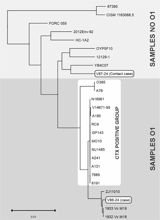
FIGURE 1: Dendrogram with strains of Vibrio cholerae O1 and non-O1, where two groups stand out (V. cholerae O1 and V. cholerae non-O1).
TABLE 2: Genomic distances based on SNPs between the reference (case strain), contact suspected case (V87/24), and the remaining isolates.
| Sample | Year | Country | SNP´s variations | Serogroup |
| Vc_M18 | 2014 | Mexico | 3846 | O1 |
| Vc_M19 | 2004 | Mexico | 3927 | |
| ZJ11010 | 2011 | China | 5895 | |
| N16961 | 1982 | Bangladesh | 14748 | |
| RC9 | 1985 | Kenya | 14775 | |
| MO10 | 1992 | India | 14963 | |
| GP143 | 1978 | Bahrein | 14993 | |
| 7685 | 2009 | Kenya | 14996 | |
| 6191 | 2005 | Kenya | 15025 | |
| MJ1485 | 1994 | Bangladesh | 15059 | |
| A131 | 1989 | India | 15088 | |
| V14671-95 | 1995 | Brazil | 15231 | |
| A241 | 1989 | Vietnam | 15252 | |
| A185 | 1992 | Colombia | 15320 | |
| A76 | 1982 | Bangladesh | 28057 | |
| O395 | 1965 | India | 28184 | |
| V87-24 | 2024 | Brazil | 47114 | Not O1 |
| YB4C07 | 2009 | USA | 49442 | |
| OYP5F10 | 2009 | USA | 50000 | |
| 12129-1 | 1985 | Australia | 50177 | |
| 2012Env-92 | 2012 | Haiti | 59313 | |
| HC-1A2 | 2010 | Haiti | 60166 | |
| FORC_055 | 2010 | South Korea | 61216 | |
| CISM_1163068.5 | 2012 | Mozambique | 116360 | |
| 87395 | 1983 | Mexico | 116642 |
SNPs (single-nucleotide polymorphisms) are single–base variations at specific positions in the genome between isolates; >40,000 SNPs indicates substantial genetic divergence, consistent with unrelated strains and no recent epidemiological linkage.
Esta reportagem reflete exclusivamente a opinião do entrevistado.