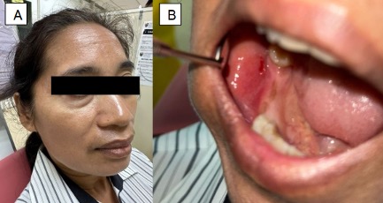
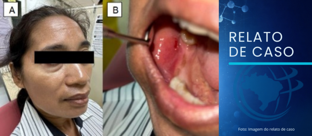
Clinical presentation of the lesion. (A). A subtle subcutaneous nodule observed over the right cheek. (B) The corresponding submucosal nodule was palpable on bimanual palpation, with mild focal erythema of the overlying right buccal mucosa
Low YK – Oral dirofilariasis of the buccal mucosa
Low Yin Kuan[1], Mohd Farifzul Anuar Mohd Zuki[1], Tan Chuey Chuan[1], WM Tilakaratne [1] and Nurul Izyan Zainuddin[1]
[1]. Universiti Malaya, Faculty of Dentistry, Department of Oral and Maxillofacial Clinical Sciences, Kuala Lumpur, Malaysia.
Corresponding author: Dr. Nurul Izyan Zainuddin. E-mail: [email protected]
Full address: Department of Oral and Maxillofacial Clinical Sciences, Faculty of Dentistry, 50603, Kuala Lumpur
Phone number: +60125647279
Conflict of Interest Statement: The author declares no conflict of interest.
Data Availability: Data is available in the body of the article.
Financial Support: There was no financial support received in the production of this manuscript.
Editor-in-Chief: Prof. Dalmo Correia Filho
ORCIDs
Low Yin Kuan: 0009-0009-8391-3326
Mohd Farifzul Anuar Mohd Zuki: 0009-0007-4365-8346
Tan Chuey Chuan: 0000-0001-6276-6202
WM Tilakaratne: 0000-0002-7061-7378
Nurul Izyan Zainuddin: 0000-0003-2035-3818
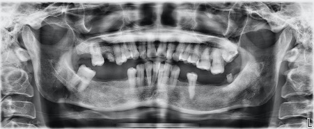
A 46-year-old Indonesian woman presented with a persistent swelling on her right cheek for one week. Clinical examination revealed a localized, well-defined firm, and movable subcutaneous nodule measuring approximately 2 2 cm (Figure 1). The nodule was also palpable intraorally through bimanual palpation. The dental panoramic radiograph (Figure 2) showed no signs of mandibular bone resorption or abnormal radiopaque or radiolucent lesions in the right mandible.
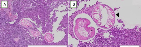
An excisional biopsy was performed, and histopathological examination revealed a soft tissue nodule with a dense infiltrate of chronic inflammatory cells, predominantly eosinophils, lymphocytes, and histiocytes. A coiled nematode, characterized by thick external cuticles with longitudinal ridges resembling a cogwheel, was identified within the fibrous connective tissue (Figure 3). The absence of uterine tubes indicated that the parasite was an adult male. A diagnosis of dirofilariasis was made, and the patient recovered without complications post-surgery.
Human dirofilariasis is a mosquito-borne zoonosis in which humans serve as dead-end hosts1. Oral involvement is rare and commonly presents as a solitary submucosal nodule resembling benign soft tissue lesions2. This rare case highlights the need to consider parasitic infections in the differential diagnosis of oral soft tissue swellings, particularly in patients from endemic areas or those in close contact with dogs. Early recognition and histopathological confirmation are crucial for accurate diagnosis and to avoid unnecessary extensive surgical procedures. Surgical excision is both diagnostic and curative.
ACKNOWLEDGMENTS
We would like to thank the institution for their support and assistance in the management of this case.
AUTHORS’ CONTRIBUTIONS
LYK: Conceptualization, Investigation, Writing – original drive; MFAMZ: Investigation; Writing – review & editing; TCC: Writing – review & editing, Supervision; WMT: Validation, Writing – review & editing; NIZ: Supervision, Validation, Writing – review & editing
REFERENCES
- Joseph M, Krishna MM, Vijayan A. Human subcutaneous dirofilariasis. Cureus. 2023;15(3):e35879. doi: 10.7759/cureus.35879.
- Jayasinghe R, Tilakaratne WM. Deep-seated lump in the buccal mucosa: dirofilariasis. In: Tilakaratne WM, Kallarakkal TG, editors. Clinicopathological correlation of oral diseases. Springer International Publishing; 2023. p. 455-62. doi: 10.1007/978-3-031-24408-7_40.